represent EMBL in Europe.
Tissue biology and disease modelling
Structural biology
Main laboratory
EMBL-EBI: European Bioinformatics Institute
Epigenetics and neurobiology
Examples: 1001, Apoferritin, Tomography, Rossmann MG, 5A1A
advanced search
Current Database
Current status
Sample type
Organism
EM Method
Resolution
Release Date
Author by name
Author by ORCID
Journal
Software
Microscope
Electron Source
Film or detector model
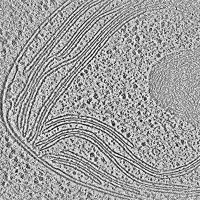
In situ cryo-electron tomogram of the Chlamydomonas chloroplast (Volta phase plate, bin4)
Wietrzynski W, Schaffer M, Tegunov D, Albert S, Kanazawa A, Plitzko JM, Baumeister W, Engel BD
eLife (2020) 9 [ DOI: doi:10.7554/eLife.53740 Pubmed: 32297859 ]
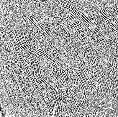
In situ cryo-electron tomogram from Chlamydomonas reinhardtii of the cellular environment around the nuclear envelope
Albert S, Schaffer M, Beck F, Mosalaganti S, Asano S, Thomas HF, Plitzko JM, Beck M, Baumeister W, Engel BD
PNAS (2017) 114 pp. 13726-13731 [ Pubmed: 29229809 DOI: doi:10.1073/pnas.1716305114 ]
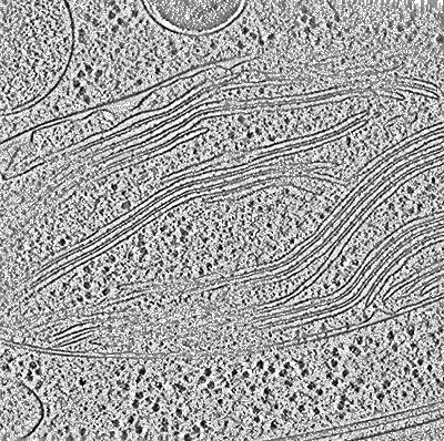
In situ subtomogram average of the Chlamydomonas double-capped 26S proteasome
Albert S, Wietrzynski W, Lee C-W, Schaffer M, Beck F, Schuller J, Salome P, Plitzko JM, Baumeister W, Engel BD
PNAS (2019) [ DOI: doi:10.1073/pnas.1905641117 ]
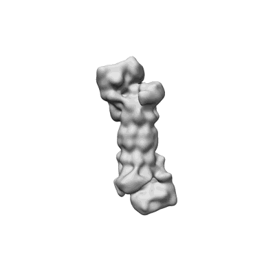
In situ subtomogram average of the Chlamydomonas single-capped 26S proteasome
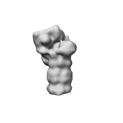
In situ subtomogram average of the Chlamydomonas ground state 26S proteasome
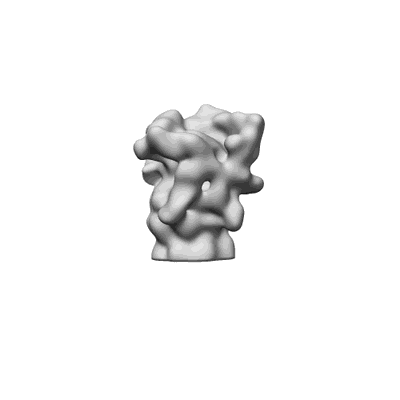
In situ subtomogram average of the Chlamydomonas processing state 26S proteasome
In situ subtomogram average of the Chlamydomonas membrane-tethered 26S proteasome
Searching in EMDBHelp
Share EMDB